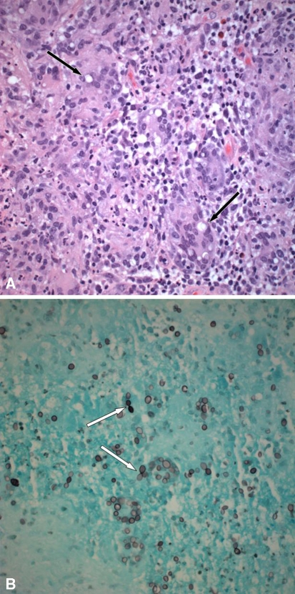
Fig. 4A–B.
(A) A representative section from formalin-fixed paraffin-embedded tissue shows an intense granulomatous reaction with numerous multinucleated giant cells containing large uninucleate yeasts (thin black arrow) (Stain, hematoxylin and eosin; original magnification, ×400). (B) A GMS stain highlights large ovoid yeasts in the cytoplasm of multinucleated giant cells. Narrow-based budding is seen with a figure-of-eight configuration (thin white arrow) (Original magnification, ×400).

