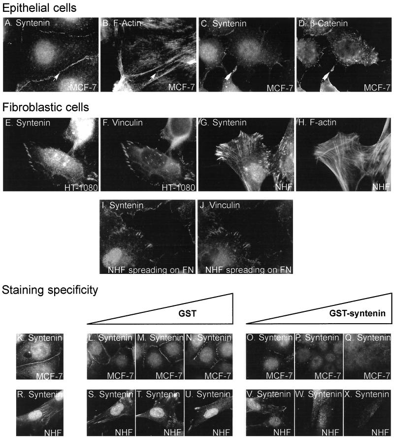
Figure 4.
Subcellular localization of endogenous syntenin. Fluorescence optical micrographs of cells cultured overnight on noncoated chamber slides (A–H and K–X) or allowed to spread for 15 min on fibronectin-coated chamber slides (I and J). The cells were stained with affinity-purified rabbit anti-syntenin polyclonal antibodies (A, C, E, G, and I) and costained with phalloidin (B and H) or with mouse mAbs specific for β-catenin (D) or vinculin (F and J). Fine arrows pinpoint colocalization of syntenin and F-actin in large cell-cell contacts, large arrows pinpoint colocalization of syntenin and β-catenin in nascent cell-cell contacts. The specificity of the stainings was addressed in competition experiments. In these experiments the cells were stained with affinity-purified rabbit polyclonal anti-syntenin antibodies (3 μg/ml) that were incubated overnight in the presence of increasing concentration of GST (0.3 μg/ml [L and S]; 3 μg/ml [M and T]; 30 μg/ml [N and U]) or GST-syntenin (0.3 μg/ml [O and V]; 3 μg/ml [P and W]; 30 μg/ml [Q and X]), or in the absence of any recombinant protein as control (K and R).

