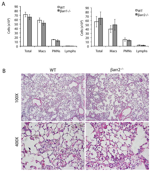
Figure 3.
Inflammatory cell influx is similar in the β-arrestin1−/−, β-arrestin2−/−, and WT mice. (A) Seven days after 1.25 U/kg bleomycin instillation, BALF cells were collected, and total cell counts as well as differential cell counts were determined. n = 5. (B) Lung sections from the β-arrestin2−/− and WT mice 7 days after bleomycin treatment were stained with hematoxylin-eosin. Original magnifications are indicated.

