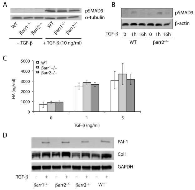
Figure 4.
Primary lung fibroblasts from β-arrestin1−/− and β-arrestin2−/− mice exhibit a normal response to TGF-β stimulation. (A–B) Fibroblasts were treated with 10 ng/ml (A) or 5 ng/ml (B) of TGF-β1 and harvested for protein after 1 (A–B) or 16 hours (B) of stimulation. Western blot analysis demonstrated a similar amount of phospho-SMAD3 in the WT, β-arrestin1−/−, and β-arrestin2−/− fibroblasts. Sample loading was verified by expression of α-tubulin or β-actin. The data are representative of three independent experiments. (C) Fibroblasts were stimulated with 1 or 5 ng/ml of TGF-β1 for 24 hours. The cell culture supernatant was removed and analyzed for HA content by ELISA. Data represent mean ± SE of four independent experiments. (D) Fibroblasts were treated with 5 ng/ml of TGF-β1 and harvested for protein after 16 hours of stimulation. Western blot analysis demonstrated a similar amount of PAI-1 and collagen I in the WT, β-arrestin1−/−, and β-arrestin2−/− fibroblasts. Sample loading was verified by expression of GAPDH.

