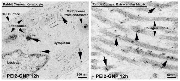
Figure 5.
Representative transmission electron microscopy images of PEI2-GNP-treated rabbit corneas demonstrating the presence and intracellular trafficking of GNP in keratoctes (A) and extracellular matrix (B). GNP can be seen in the endosomes near the cell surface depicting their uptake by endocytosis. Ruptured endosome represents the release of GNP into the cytoplasm.

