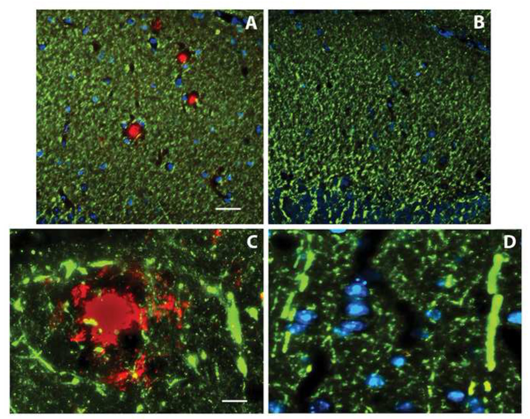
Figure 2.
Loss and dysmorphology of dendrites is confined to amyloid plaques. MAP2 immunostaining (green) of the dentate molecular layer (A,B) and parietal neocortex (C,D) of the 14 month old DKI mouse either not receiving treatment (A,C) or six months after initiating Aβ vaccination (B,D). Panel A illustrates that the dense dendritic network is interrupted by spheres that immunolabel for Aβ (red). Neither amyloid deposits nor gaps in the dendritic network are observed following Aβ vaccination (B). Nuclei labeled with DAPI are shown in blue. At high magnification, amyloid deposits are surrounded by curvy dendrites (C). This abnormal dendritic morphology is not found following Aβ vaccination (D). Scale bar= 50 µm (A,B); 10 µm (C,D).

