
FIGURE 15.
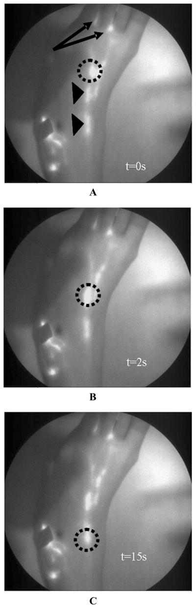
White-light overlay with corresponding fluorescent images of lymphatic map of foot of a 46-year-old female who was injected with 100 μL of 25 μg of IC-Green in the first and second interdigital spaces (arrows) in the dorsum of the foot. Circles indicate a “packet” of dye transiting along the lymphatic channel. Arrowheads identify localized collection of fluorescent dye that could be lymphangions of a lymphatic vessel. Panels A, B and C depict images at 0,2, and 15 s.