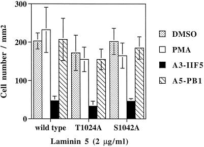
Figure 6.
Cell adhesion of CHO-α3 transfectants. Cells were tested for adhesion to laminin-5 (coated at 2 μg/ml), and attached cells were analyzed by using the Cytofluor 2300 measurement system (Millipore, Bedford, MA) as previously described (Chan et al., 1992; Bazzoni et al., 1995). Cells were untreated, incubated with 100 nM PMA at the start of the assay, or were preincubated with either antihuman α3 (A3-IIF5) or anti-hamster α5 (PB1) mAbs at 4°C for 30 min before the adhesion assay. Background binding (assessed by using bovine serum albumin-coated wells) was typically less than 5% of the total and was subtracted from experimental values. Results are reported as mean ± SD of triplicate determinations. This experiment was repeated multiple times, with each experiment yielding no significant differences between mutant and wild-type α3 transfectants.

