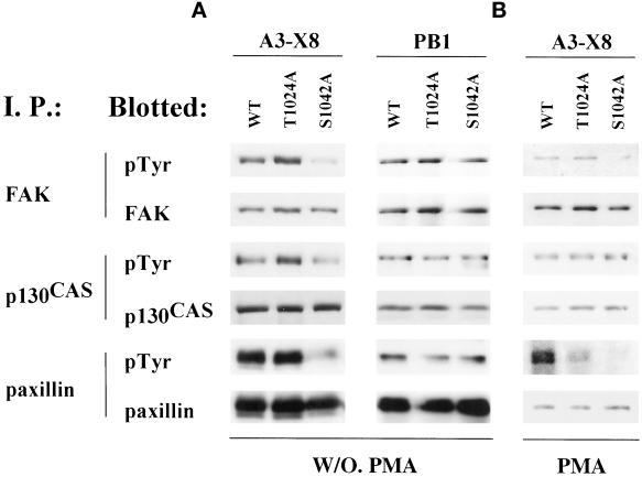
Figure 7.
Signaling through wild-type and mutant integrin α3. (A) After detaching, washing, and resuspending in serum-free MEMα+ media, CHO-α3 transfectant cells were allowed to spread on surfaces coated with either anti-human α3 mAb X8 (10 μg/ml) or anti-hamster α5 mAb PB1 (10 μg/ml) at 37°C in10% CO2 for 60 min. Cells were then lysed in RIPA buffer, and immunoprecipitations (I. P.) were carried out using antibodies to the indicated proteins. After SDS-PAGE under reducing conditions, proteins were transferred to nitrocellulose membranes, and blotted with anti-phosphotyrosine mAb, and then stripped and reblotted with antibodies to the indicated proteins. (B) CHO transfectants were treated with 100 nM PMA for 30 min before lysis, and then analyzed as in A.

