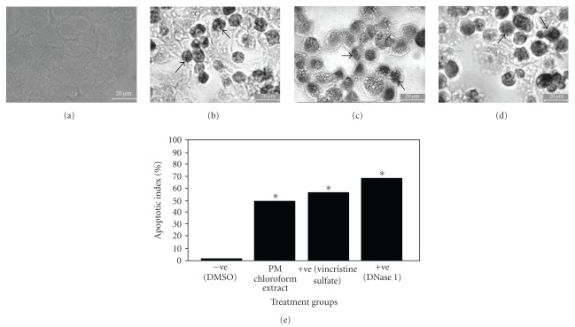
Figure 2.
The effect of (a) DMSO l% (v/v), (b) Physalis minima chloroform extract (2.80 μg/mL, EC50 a 72 h), (c) DNase I (1 U/mL) and (d) vincrstine sulphate (0.0015 μg/mL, EC50 at 72 h) or NCI-H2: cells for 24 h and subjected to Deadend Colometric Apoptosis Detection System (Promega, USA) Apoptotic cells with stained nuclei were rnarked by arrows. (e) Comparison of the mean percentage of apoptotic index between DNase 1-, vincristine sulphate- and P. minima chloroform extract-treated cells to untreated cells (DMSO) at 24 hours treatment in different cell lines. Each value represented mean ± SEM from three independent experiments. *P < .05 (as compared with negative control).

