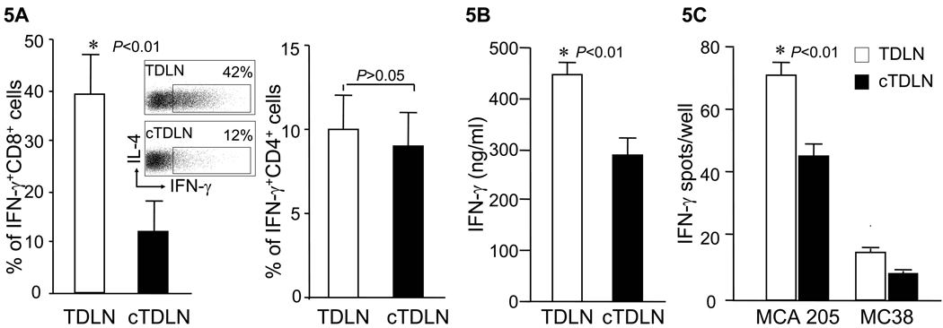
Figure 5.
cTDLN cells produced less IFNγ compared to TDLN cells. A. Percent of IFNγ+CD8+ cells was lower in activated/expanded cTDLN compared to TDLN, while the percent of IFNγ+CD4+ cells was similar in TDLN and cTDLN. The percent of IFNγ+ T cells was determined by intracellular staining and analyzed by FACS analysis, gated on CD8+CD90+ and CD4+CD90+ T cells. Results were expressed as the mean percent of IFNγ+ T cells ± SEM (8 independent experiments). B. The secretion of IFNγ was lower in expanded cTDLN compared to TDLN. Supernatants were collected from the expanded cTDLN and TDLN cells. IFNγ was detected by ELISA. Results were expressed as the mean values of IFNγ ± SEM of triplicate samples from 3 independent experiments. C. The frequency of tumor specific IFNγ-producing effector cells was significantly less in the cTDLN population. 2×10⁵ effector cells were cultured alone or in the presence of 5×10⁴ irradiated specific MCA 205 or irrelevant mouse MC38 tumor cells for 24hr in 96-well ELISPOT plates coated with anti-IFN-γ. The number of spots from wells containing effector cells alone was subtracted from the corresponding wells for each sample to determine the number of tumor-induced spots. Results were expressed as the mean number of spots/well ± SEM. (5 independent experiments).

