
Figure 4. Prom1−/− cells have increased proliferation in vitro.
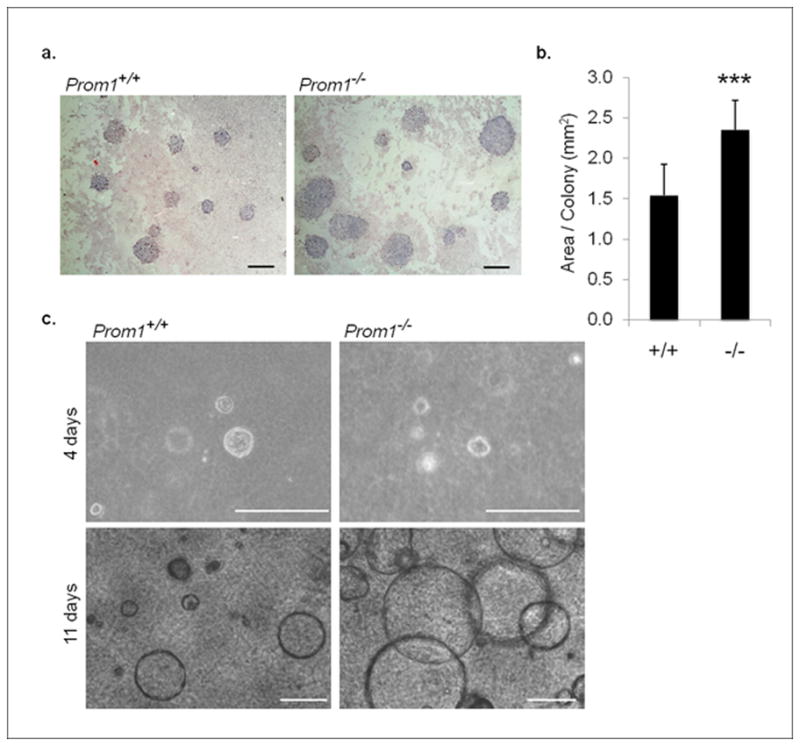
(a) Giemsa-stained 2D culture assays, revealing colonies derived from FACS-sorted epithelial cells; scale bars: 2 mm. (b) Quantification of the area of individual colonies from Prom1+/+ and Prom1−/− cells (n=12). (c) Spherical colonies from Prom1+/+ and Prom1−/− mammary epithelial cells in 3D culture, shown after 4 days and 11 days of growth; scale bars: 200 μm. ***p<0.001, bars=SD.