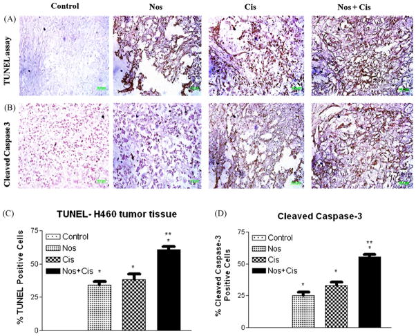
Fig. 6.
Immunohistochemical staining of xenograft H460 lung tumor tissues for induction of apoptosis using TUNEL assay (A); for expression of cleaved caspase 3 (B); quantitation of apoptotic cells from TUNEL staining (C); and quantitation of cleaved caspase 3-positive cells apoptotic cells (D). Tumor tissues were dissected from mice on day 38, fixed in 10% formalin, paraffin embedded and sectioned. Sections were stained using the DeadEnd colorimetric kit and cleaved caspase 3 (Asp 175) IHC kit for TUNEL assay and cleaved caspase 3 expression as described in Section 2 respectively. The apoptotic tumor cells are stained brown. Percentages of TUNEL-positive and cleaved caspase 3-positive cells were quantitated by counting 100 cells from 6 random microscopic fields. Data are expressed as mean + SD (N = 6). One-way ANOVA followed by post-Tukey test was used for statistical analysis to compare control and treated groups. P < 0.01 (*significantly different from untreated controls; **significantly different from Nos and Cis single treatments). Original magnification 40× (micron bar = 50 μm).

