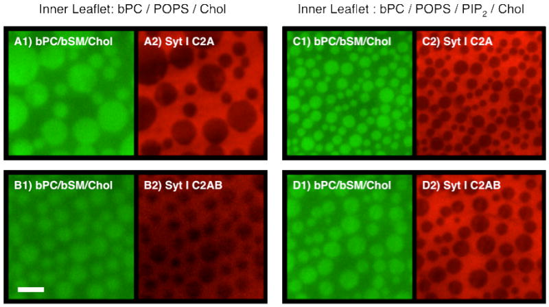
FIGURE 2.

Representative images of Ca2+-mediated synaptotagmin I C2A and C2AB domain binding to asymmetric two-phase bilayers with outer leaflet monolayers composed of bPC/bSM (1:1) plus 20% cholesterol and supported on a polymer cushion and inner leaflet monolayers composed of bPC, 15% POPS and 20% cholesterol (left panels) or bPC, 15% POPS, 1% bPIP2 and 20% cholesterol (right panels). The outer leaflet monolayers are visualized with 0.25% NBD-DPPE, which preferentially partitions into lo phases (green images). The inner leaflet monolayers are not labeled, but are known to form induced lo phases on top of outer leaflet lo phases (30). Alexa-546 labeled synaptotagmin I C2A (top panels) and C2AB (bottom panels) domains bind to induced inner leaflet lo and ld phase domains, and preferentially partition to the more disordered lipid phase regions (red images). Scale bar: 10 μm.
