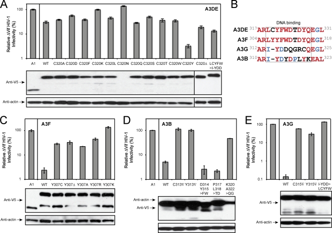
Fig. 2.
Characterization of A3 DNA binding domain. The anti-HIV-1 activity of A3DE mutants (A), A3F mutants (C), A3B mutants (D), and A3G mutants (E) were determined as in Fig. 1. Lower panels show protein expression in viral producer cells, which were determined by Western blotting with the indicated antibodies. (B) Amino acid sequence alignment of A3B, A3DE, A3F, and A3G regions, where the A3DE C320 residue is located. Completely conserved, partially conserved, and nonconserved residues are shown in red, blue, or black, respectively. WT, wild type.

