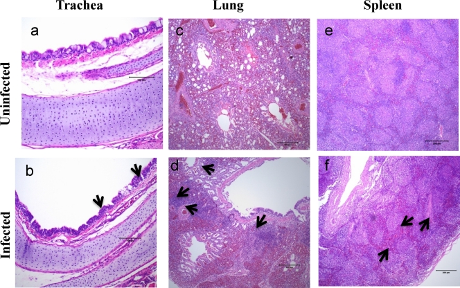
Fig. 5.
Histopathology in sections of trachea, lung, and spleen harvested from 2-week-old chickens 3 days after inoculation with parental rAPMV-2 or the rAPMV-2 (type 4), rAPMV-2 (type 3), rAPMV-2 (type 1v), or rAPMV-2 (type 5) mutant by the oculonasal route. Chickens were mock infected (a, c, and e) or infected with parental rAPMV-2 virus (b, d, and f). Histopathology in sections of chicken tissues infected with mutant viruses looked similar to that for tissues infected with parental virus. Sections were stained with hematoxylin and eosin (magnification, ×400). Histopathological examinations of tissue samples revealed similar microscopic findings in all tested recombinant viruses. (b) In the infected trachea, minimal to mild attenuation and flattening of the tracheal epithelium, with reduction and loss of cilia, were observed. There was loss of normal columnar epithelial architecture in these regions, with mild epithelial hyperplasia and multifocal replacement by low cuboidal epithelial cells (arrows). Low numbers of individually apoptotic cells were seen within the epithelium in these regions. There was mild, multifocal, subepithelial infiltrate of lymphocytes and fewer macrophages in the lamina propria. In summary, minimal to mild, multifocal lymphocytic tracheitis with epithelial attenuation and regeneration was noted. (d) In the infected lung, small to moderate numbers of lymphocytes and few macrophages were seen infiltrating around blood vessels and within the interstitium. Inflammatory cells formed dense perivascular aggregates that extend into the interstitium, with small numbers of inflammatory cells multifocally infiltrating into the lamina propria subjacent to the airway epithelium. Small numbers of individually apoptotic cells were present in inflammatory aggregates. Mild to moderate, multifocal, lymphohistiocytic, perivascular and interstitial pneumonia was observed. (f) In the infected spleen, the periarteriolar sheaths and white pulp regions exhibited minimally reduced numbers of lymphocytes. There was also minimal lymphoid depletion.

