Fig. 1.
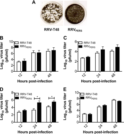
Differential plaque morphology and growth kinetics of RRVPERS compared to those of RRV-T48. (A) Plaque morphology on Vero cell monolayers for parent Ross River virus strain T48 (RRV-T48) and RRVPERS. Both RRV-T48 and RRVPERS plaques were purified from the supernatants of RRV-T48-infected RAW 264.7 cultures at day 14 postinfection, and new viral stocks were regrown in fresh Vero cell cultures, after which plaque morphology was confirmed by plaque assay. (B and D) Growth kinetics of RRV-T48 and RRVPERS in Vero (B), HEp-2 (C), or RAW 264.7 (D) cells infected at an MOI of 0.1. (E) RAW 264.7 cells were pretreated with 104 U/ml of anti-murine IFN-α and anti-murine IFN-β antibodies (R&D Systems) for 1 h at 37°C. The cells were then infected with 0.1 MOI RRV-T48 or RRVPERS for 1 h at 37°C. The virus inoculum was discarded, and cells were rinsed with medium. Fresh medium containing 104 U/ml of anti-murine IFN-α and anti-murine IFN-β antibodies was added to the monolayer for an additional 48 h at 37°C. Culture supernatants were collected at various time points post-RRV infection, and viral growth was assessed by plaque assay on Vero cell monolayers. The assay limit of detection is 2.0 log10 PFU/ml. Significant differences in virus titers (P < 0.05) are marked with an asterisk.

