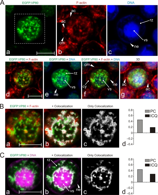
Fig. 6.
Colocalization of VP80 with F-actin filaments connecting the virogenic stroma with the nuclear envelope. (A) Confocal microscopy of Sf9 cells infected with Ac-Δvp80-egfp:vp80 (MOI = 10) expressing EGFP-fused VP80 (green). At 24 h p.i., the cells were fixed and stained with Hoechst 33342 for DNA (blue) and with phalloidin-rhodamine for F-actin (red). The images were collected in green (a), red (b), and blue (c) channels and differentially merged (d to f), and, finally, 3D reconstructions of confocal Z-stack data sets were performed (g). (B) Colocalization of EGFP-VP80 with nuclear F-actin. (C) Colocalization of EGFP-VP80 with DNA-labeled regions. For panels B and C, the green and red channels from the inset from panel Aa were merged (a). Colocalization regions are depicted in white (b to c). (B and C) The colocalization proportions as represented by PC and ICQ values were plotted into graphs (d). Abbreviations: ac, cytoplasmic F-actin cables; cm, cytoplasmic membrane; cy, cytoplasm; ne, nuclear envelope; rz, ring zone; vs, virogenic stroma. Scale bars represent 10 μm.

