Fig. 3.
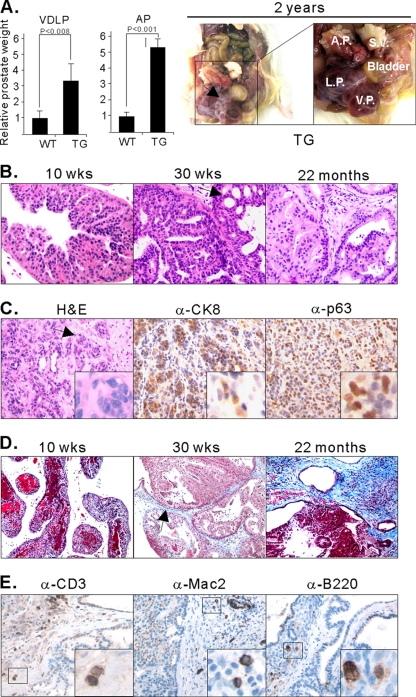
The lesions induced by MT resulted in severe phenotypes, invasion, and reactive stroma. (A) Enlargement of prostate lobes of TG mice. The weights of the VP/DLP (VDLP) were combined to avoid possible errors during tissue isolation. The weights of prostate tissues were normalized with the body weights. The graph was then standardized to the weight of VP/DLP of the wild type, and the relative differences between the weights of tissues from wild-type and transgenic mice are presented. Error bars show standard deviations. Prostates of WT mice were obtained from mice 91 to 92 weeks old, and prostates of TG mice were isolated from mice 81 to 96 weeks old. Frank tumors developed, as seen in an approximately 2-year-old FF23 line mouse (right panels). S.V., seminal vesicle. (B) H&E staining of DLP isolated from GG982 line mice at 10 weeks, 30 weeks, and 2 years of age. The black arrow indicates the cribriform phenotype. At least 5 mice were examined at each specific age. (C) H&E staining of the AP of a 2-year-old FF23 line mouse exhibits extensive invasive prostate cancer. IHC assays were conducted with anti-CK8 and anti-p63 antibodies. The black arrow indicates irregular nests of atypical epithelial cells penetrating the stroma surrounding the prostatic ducts, indicating invasion. (D) Masson's trichrome staining shows the development of reactive stroma in blue color in old mice (>30 weeks old). The black arrow indicates blue staining with Masson's Trichrome. (E) IHC experiments with anti-CD3, anti-B220, and anti-MAC2 antibody show infiltration of inflammatory cells.

