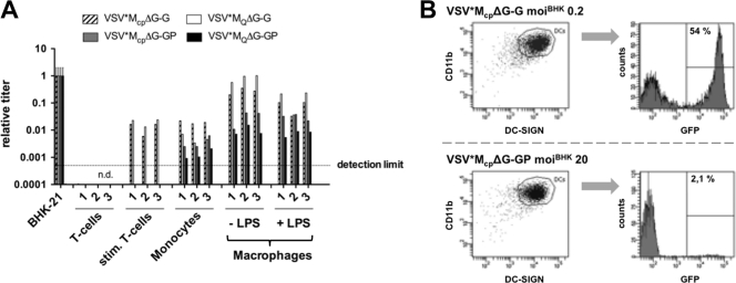
Fig. 2.
VSV*ΔG-GP shows negligible transduction of PBMCs, macrophages, and dendritic cells. PBMCs were isolated from blood and T cells were stimulated as previously described (30). CD14+ monocytes were purified by magnetic sorting using anti-CD14 microbeads (Miltenyi Biotec). CD14+ monocyte-derived macrophages were differentiated in vitro by adding 50 ng/ml recombinant human macrophage colony stimulation factor (rhM-CSF) (Invitrogen) for 6 days. Monocyte-derived DCs were generated after culturing CD14+ cells with IL-4 and GM-CSF in vitro (35). Macrophages were activated overnight by LPS stimulation (100 ng/ml). Freshly isolated T cells and activated T cells as well as monocytes and macrophages (A) and DCs (B) were infected with 10-fold serial dilutions of VSV*ΔG-G and VSV*ΔG-GP. Titers were analyzed by flow cytometry based on the expression of GFP and normalized to the reference BHK-21 hamster cell line. GFP expression by DCs (gated on CD11b+/DC-SIGN+) was quantified 16 h postinfection by flow cytometry. n.d., not detectable.

