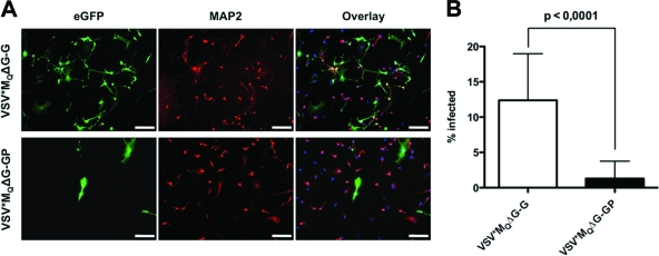
Fig. 3.
Reduced neurotropism of LCMV-GP pseudotypes in vitro. Primary human neurons (ScienCell Research Laboratories) were grown on 2 μg/cm2 poly-l-lysine-coated culture vessels and infected with VSV*MQΔG-G and VSV*MQΔG-GP at an MOIBHK of 4 (A) or 1 (B). At 16 h postransduction, cultures were fixed in 4% paraformaldehyde. Neurons were stained with mouse anti-MAP2 (Sigma) primary antibody and goat anti-mouse Alexa Fluor 568-conjugated (Invitrogen) secondary antibody. Nuclei were counterstained with DAPI (Roche). (A) A representative microscopic field is shown. eGFP, enhanced GFP. Bars, 200 μm. (B) The percentages of GFP+/MAP2+ cells were determined by fluorescence microscopy, counting 30 microscopic fields per pseudotype. Values are expressed as means ± standard deviations (SD) and were tested for significance by Student's t test (P < 0.0001).

