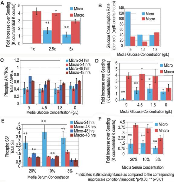
Fig. 3.
Volume density and media supplementation assays. Volume density changes alone were not sufficient to increase microchannel proliferation, and in both 2.5× and 5× densities (1× density being the typical macroscale volume density), proliferation was significantly reduced as compared to macroscale cultures of the same density (A). Glucose consumption rates in media with a dilution of starting glucose concentration were assayed (B). ICWs for AMPK phosphorylation at 24 and 48 h after seeding for macro- and microscale cultures with the dilution of glucose showed no significant differences between any of the conditions (C), though proliferation was significantly reduced in microchannels, except for the 0 g L−1 condition (D). Significant differences between microchannel cultures and the corresponding macroscale culture at the same time points were seen regardless of media serum concentrations (E), though proliferation was significantly lower in microchannel cultures regardless of media serum concentration (F). All proliferation data listed here compare the fold increase in cell number at 48 h versus that at 3 h post-seeding.

