Figure 5.
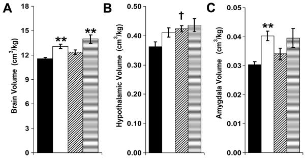
Magnetic resonance imaging was performed to assess male mice for neonatal GR or leptin-induced alterations in regional brain volumes (■: control-saline, n=15; □: GR-saline, n=12; ▨: control-leptin, n=8; ▤: GR-leptin, n=6). Compared to control mice, GR mice had increased brain volume to body weight ratios (A; F(1,37)=26, **P<0.01 versus corresponding control). Although neonatal leptin administration also increased relative brain volumes (A; F(1,37)=7.3, P=0.01), no significant pair-wise differences were present (P=0.06 for control-leptin versus control-saline and P=0.07 for GR-leptin versus GR-saline). Relative hypothalamic volumes were significantly increased by neonatal leptin supplementation (B; F(1,37)=5.1, †P=0.02 for control-leptin versus control-saline). Neonatal GR significantly increased relative amygdala volumes (C; F(1,37)=14, **P<0.001 GR-saline versus control-saline, P=0.11 GR-leptin versus control-leptin).

