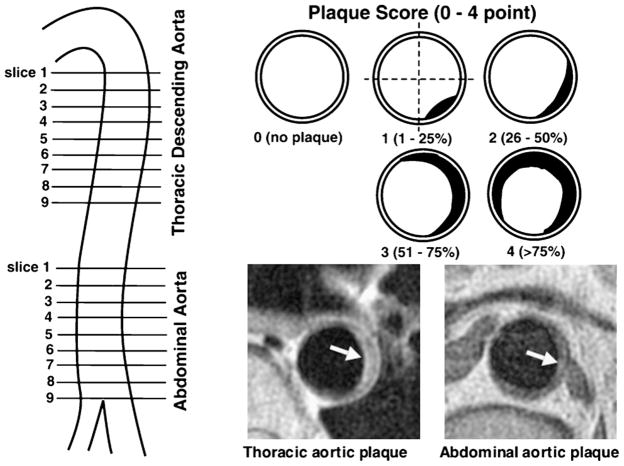
Figure 1.
MRI slices of the aortas and plaque scores. For each patient, 9 slices of thoracic descending aorta and 9 slices of abdominal aorta were obtained at 12-mm intervals, which each covered about 10-cm portion of thoracic aorta below aortic arch and 10-cm portion of abdominal aorta above the bifurcation of iliac artery. Plaque was defined as a clearly identified luminal protrusion with focal wall thickening, and plaque extent in each slice was scored from 0 to 4 points by the percentage of luminal surface involved by plaque. Arrows indicate aortic plaques.

