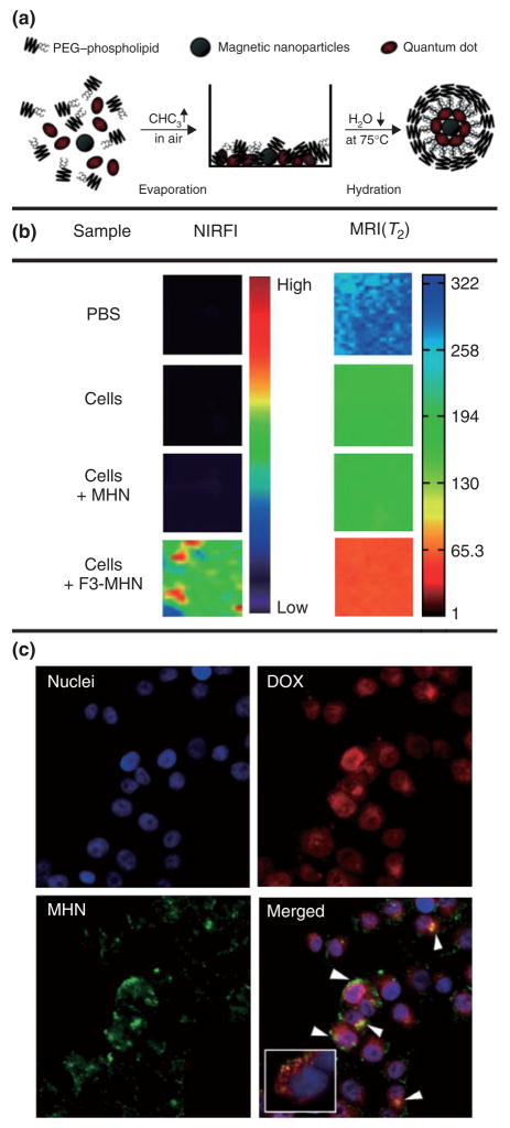
FIGURE 7.
(a) Synthetic procedure used to prepare micellar hybrid nanoparticles that encapsulate magnetic nanoparticles and quantum dots within a single PEG-modified phopholipid micelle. (b) Multimodal images (NIR fluorescence in the Cy5.5 channel and MRI) of PBS and MDA-MB-435 human carcinoma cells that were left untreated, were treated with untargeted nanoparticles and with targeted nanoparticles. (c) Targeted drug delivery of nanoparticles containing DOX into MDAMB-435 human carcinoma cells. The DOX-loaded nanoparticles were incubated with the cells for 2 h. Arrowheads indicate colocalization of DOX and nanoparticles. The inset shows the colocalization of some DOX (red) and the endosome marker (green) 30 min after incubation with DOX-loaded nanoparticles. The nuclei were stained with 4–6-diamidino-2-phenylindole. Reproduced with permission from the publisher.54

