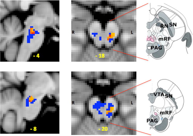
Figure 3.
Opioid withdrawal-induced effects on neuronal response (BOLD activity) within the MPRF. The effect of opioid withdrawal on neuronal response (BOLD activity) to thermal noxious stimulation (X) was defined by (Xpostinfusion (opioid) − Xpreinfusion(opioid)) − (Xpostinfusion (saline) − Xpreinfusion(saline)). The left sagittal (left column) and axial (center column) slices show the cluster of significantly increased activity (red/yellow) induced by opioid withdrawal. MNI coordinates (in yellow) are given at the bottom of each slice. The small volume correction was based on the MPRF region of interest (blue voxels). The annotated diagrams on the right are drawings of the axial views of the brainstem to aid visual localization of the area of increased activity within the MPRF [adapted from Duvernoy (1995)]. VTA, Ventral tegmentum area; SN, substantia nigra; RN, red nucleus; mRF, mesencephalic reticular formation.

