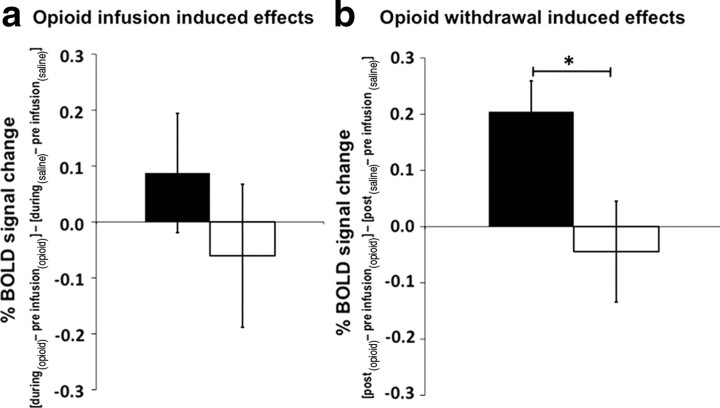
Figure 4.
a, b, Opioid infusion-induced effects (a) and opioid withdrawal-induced effects (b) on neuronal response to thermal noxious stimulation within the isolated MPRF in responders (black bars) and nonresponders (open bars). The y-axes show the induced effects on neuronal response as percentage BOLD signal change. Opioid infusion-induced effect on neuronal response (X) was defined by (Xduring-infusion (opioid) − Xpreinfusion (opioid)) − (Xduring-infusion (saline) − X preinfusion (saline)). Opioid withdrawal-induced effect on neuronal response (X) was defined by (Xpostinfusion (opioid) − Xpreinfusion(opioid)) − (Xpostinfusion (saline) − Xpreinfusion(saline)). Error bars indicate SEM. (*p < 0.05). The opioid withdrawal-induced neuronal response to thermal noxious stimulation in the MPRF is significantly higher in responders than in nonresponders. The effect of the opioid infusion on BOLD activity in the MPRF appears to increase in responders and decrease in nonresponders, but these effects are not statistically significant.

