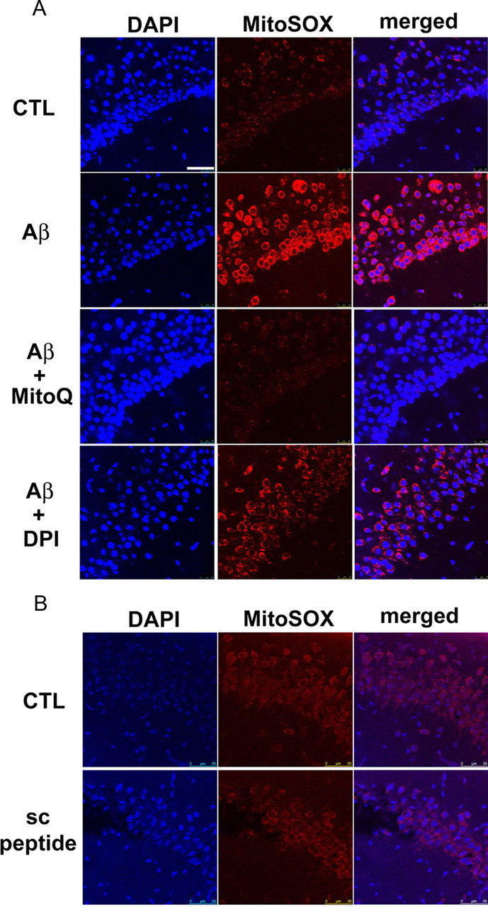
Figure 3.

MitoQ, but not DPI, prevents Aβ-induced increases in mitochondrial superoxide. A, Treatment of hippocampal slices with Aβ1-42 (500 nm) for 60 min increased the MitoSOX fluorescent signal (red) in area CA1 compared with control slices. DAPI staining is shown as blue. Pretreatment of slices with MitoQ (500 nm) blunted the Aβ-induced elevation in the MitoSOX fluorescent signal. In slices pretreated with DPI (10 nm), Aβ still caused increases in the MitoSOX fluorescent signal. B, Treatment of slices with a scrambled Aβ1-42 peptide (500 nm) for 60 min did not alter the MitoSOX fluorescent signal. Results are representative of three independent experiments. Scale bar, 50 μm. CTL, Control; sc, scrambled.
