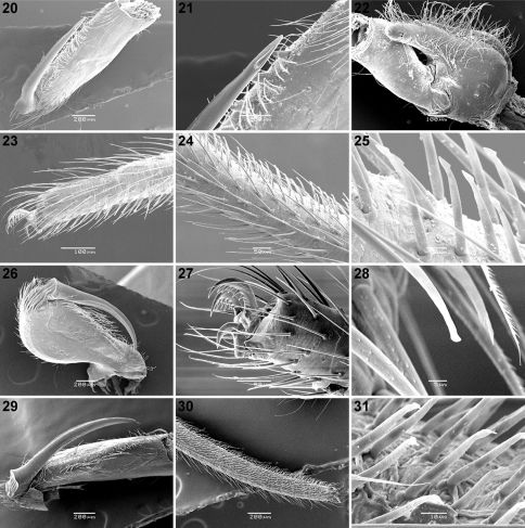
Figures 20–31.
Scanning electron micrographs of Calommata meridionalis sp. n. 20–25 Calommata simoni Pocock 26–28 and Calommata tibialis sp. n. 29–31 males 20, 29 chelicera in ventral view 21 tip of fang 22 right endite, ventral view 23, 27 tarsus and claw, leg I (note pseudosegmentation of tarsus) 24, 30 tarsus IV, lateral and ventral view (note pseudosegmentation of tarsus) 25, 28, 31 detail of ventral scopulate setae on tarsus IV 26 chelicera in prolateral view.

