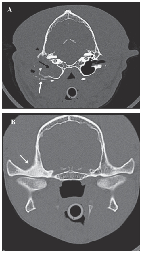
Figure 1.
A — Transverse slice obtained at the level of the ossicular chain shows, at bone setting, complete loss of air contrast associated with soft tissue density within the middle ear cavity (black arrow); enlargement of the tympanic cavity and bulla with mild osteolysis and sclerosis of the latero-ventral aspect of the bulla (white arrow), and sclerosis of the temporal bone (arrowhead); absence of air contrast in the horizontal tract of the ear canal (compared with contralateral ear canal); disappearance of the ossicular chain and mild erosion of the promontory. B — Transverse slice obtained at the level of the TMJ at bone setting; note the severe osteosclerosis of the right mandibular fossa (white arrow).

