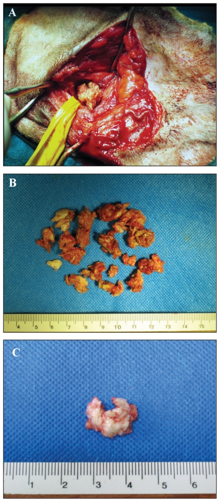
Figure 3.
Gross appearance of middle ear cholesteatoma during surgery. A — Spongy aspect of the keratinous material; the material spontaneously protruded after enlargement of the bulla opening; B — All material removed from the middle ear cavity in the same ear, note the keratinous appearance; C — Cyst-like aspect of cholesteatoma after surgical removal.

