Figure 4.
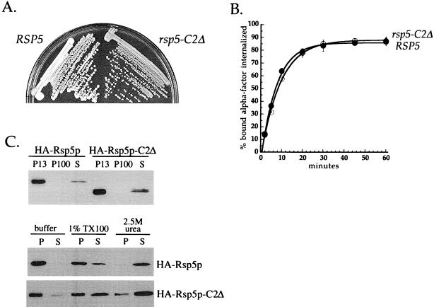
Phenotypic analysis of rsp5-C2Δ cells. (A) Growth of RSP5 and rsp5-C2Δ cells. LHY1103 (RSP5) and LHY1101 (rsp5-C2Δ) cells were streaked onto YPUAD medium and grown for 2 d at 30°C. (B) α-Factor internalization assays performed by the pulse-chase protocol at 30°C (see MATERIALS AND METHODS). Cells were propagated in SD medium at 30°C. RSP5 (LHY1103, ●), rsp5-C2Δ (LHY1101, ○). (C) Fractionation of lysates prepared from cells expressing HA-Rsp5p or HA-Rsp5p-C2Δ. Top, differential centrifugation of LHY2066 (HA-Rsp5p) and LHY2232 (HA-Rsp5p-C2Δ) lysates. Cells were propagated in casamino acids-galactose medium at 24°C. Fractionation and immunoblot analysis were performed as described for Figure 3A. Bottom, fractionation of LHY1098 (HA-Rsp5p) and LHY1101 (HA-Rsp5p-C2Δ) cell lysates. Cells were propagated in YPUAD medium at 30°C. Lysates were fractionated into 100,000 × g pellet and supernatant fractions after biochemical treatment with 1.0% Triton X-100 (TX100), 2.5 M urea, or buffer as described for Figure 3.

