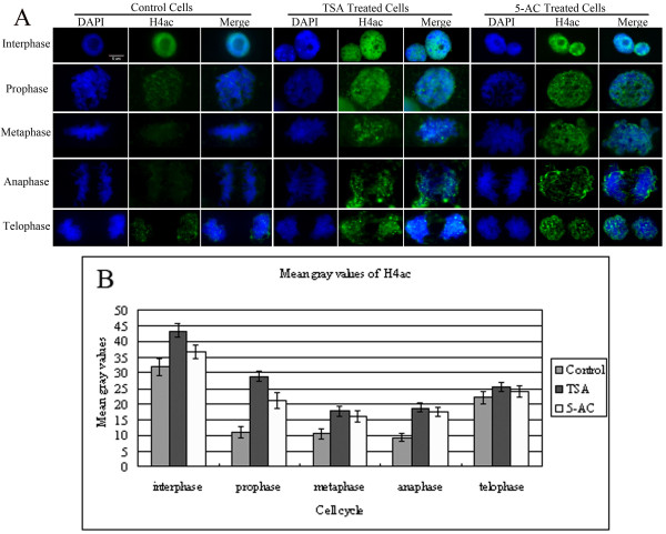
Figure 4.
Increased levels of H4ac in TSA- and 5-AC-treated cells. (A) Immunostaining of H4ac in representative maize root tip cells in interphase or mitosis after TSA or 5-AC treatment. The 'DAPI' panel shows DAPI-stained DNA images, the 'H4ac' panel shows immunostained images and the 'Merge' panel shows the combined blue and green signals. In control cells, the strongly acetylated histone H4 signal was evenly distributed in the nucleus at interphase, and nucleoli were barely acetylated. At prophase, the deacetylation of H4 is initiated, and very weak acetylation is observed. As the cells progressed into metaphase and early telophase, the acetylated histone H4 signal is almost undetectable, whereas at telophase, H4 begins to be acetylated. In both TSA- and 5-AC-treated cells, strong hyperacetylation of histone H4 was detected in both mitotic and interphase cells of maize root tips compared with control cells. Bar = 5 μm. (B) Histogram showing mean gray values of the immunostaining signals for H4ac shown in (A). The mean gray value for H4ac after treatment with either TSA or 5-AC is higher than in the control. The mean gray values for H4ac were showed in Additional file 1: Table S1. Error bars represent the standard error of the mean.

