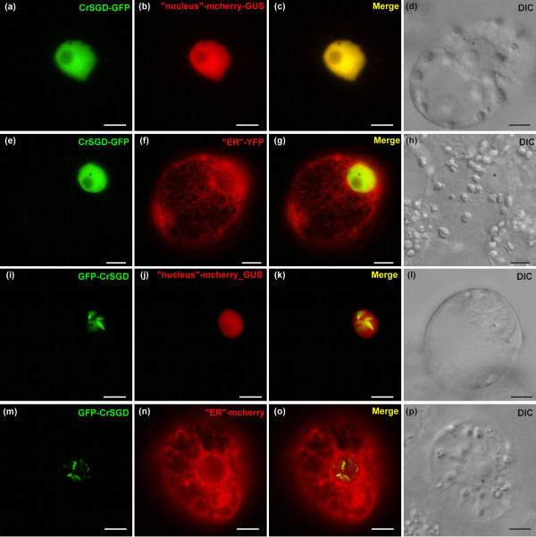
Figure 6.
CrSGD is localised to the nucleus of C. roseus cells with a diffuse or a punctuated/fusiform aggregation-like pattern of fluorescence dependant on the orientation of the GFP fusion. Undifferentiated C. roseus cells were co-transformed with CrSGD-GFP or GFP-CrSGD constructs (1st column) together with mcherry or YFP organelle markers constructs (2nd column). The merged image and the DIC morphology are presented in the 3rd and 4th columns, respectively. Note that SGD fusions were localised to the nucleus and not to the ER with either a diffuse fluorescence pattern (CrSGD-GFP) and a punctuated or fusiform aggregation-like pattern of fluorescence (GFP-CrSGD). Bar: 10 μm.

