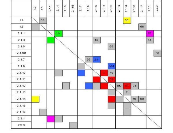
Figure 1.

Overview of microarray analyses performed. Tissues are specified by their IDs (see table 2 for details). Numbers specify the amount of differentially expressed genes in an experiment (p ≤ 0.005). Those experiments that are discussed in detail are marked with colours according to the experimental question: development of somatic embryos (red), putative reasons for developmental arrest in the globular stage (blue), comparison of embryogenic and non-embryogenic cell cultures (magenta), comparison of somatic and zygotic embryos (yellow) as well as comparison of a diploid and a tetraploid callus line (green).
