Figure 3.
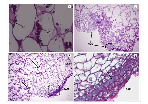
Histological analysis of embryogenic/organogenic callus of V. planifolia on A4 medium and A10 medium 20 days after subculture. Four-micrometer-thick sections of callus were stained with Periodic acid and Schiff' reagent. a. Staining for starch revealed cell amyloplasts (CA) in vacuolated large cells of CA10 d20 callus inner region; bar = 20 μm. The histological section from CA4 d20 showed the same figure. b. Nodular compact structure (NCS) containing small mitotic cell in the peripheral region of CA4 d20 callus and indicating PLB early formation; bar = 100 μm. c. Apex shoot differenciation in peripheral region of CA10 d20 callus: development of shoot apical meristem (SAM) and leaf sheath primodium (LSP). Cell amyloplasts (CA) near the site of shoot formation can be seen in large vacuolated cells; bar = 100 μm. d. Higher magnification of shoot apical meristem (SAM) in CA10 d20: cell layers in tunica development are distinguished (arrow); bar = 25 μm.

