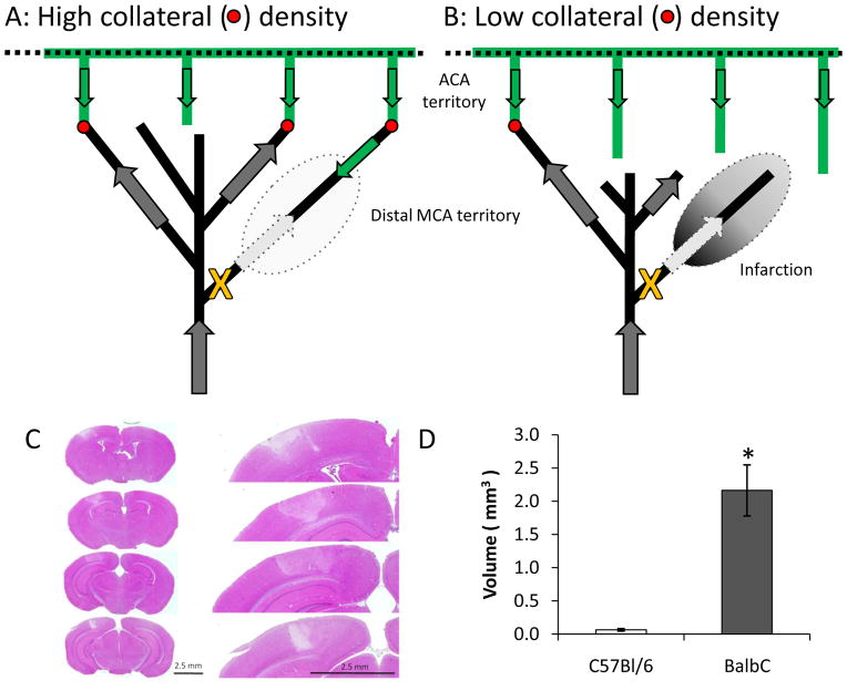
Figure 10.
Schematic illustrating the differences between C57Bl/6J and BALB/c collaterals and their impact on post-occlusion blood flow in the distal MCA branch. (C) Hematoxylin and eosin staining from the BALB/c mouse. Minimal infarction was observed in the C57BL/6J and was constrained to the site of irradiation. (D) Quantification of infarct volume (* p<0.02 two-tailed heteroscedastic t-test, N=6 C57BL/6J and 4 BALB/c mice). One BALB/c mouse showed minimal infarction and was excluded. Inclusion of this mouse decreased significance to p<0.04.

