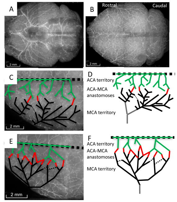
Figure 2.
Vessel painting reveals strain-specific variation in pial vessel architecture at six months of age. (A) Ventral view of a BALB/c mouse brain after vessel painting. (B) Dorsal view of the cortical pial vessels. (C & D) Tracing the major ACA (green) and MCA (black) vessels and their connections (red) in the BALB/c mouse. (E & F) Increased collateral density in C57BL/6J mice.

