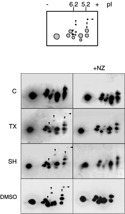
Figure 2.
MT assembly results in at least four major specific phosphorylated stathmin/Op18 forms. Two-dimensional gel analysis of high-speed mitotic Xenopus egg extract supernatants (immunoblots probed with antiserum I) shows four extra spots (arrowheads) in conditions where MT assembly was induced (TX, SH, DMSO). These spots were absent in the same conditions with 10 μM NZ (right). Schematic representation of the stathmin/Op18 phosphorylation pattern: spots present in control and nocodazole-containing fractions are gray, the additional spots induced by MT assembly are black.

