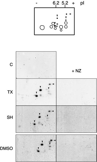
Figure 4.
MT assembly-dependent phosphorylation on Ser 16. Two-dimensional immunoblots of Xenopus high-speed mitotic egg extracts probed with antiphosphorylated Ser 16 stathmin/Op18 antiserum (anti-Ser16P). In all conditions where MTs were allowed to polymerize for 45 min, four main spots (arrowheads) corresponding to hyperphosphorylated isoforms revealed by antiserum I were detected (Figure 2). Several other minor spots were detected as well. They were not observed when nocodazole (NZ) was present. Schematic representation of the total stathmin/Op18 phosphorylation pattern as found in Figure 2 is shown on top: the additional spots induced by MT assembly and recognized by anti-Ser16P are black and pointed by arrowheads; spots present in control and nocodazole-containing fractions are not recognized by anti-Ser16P.

