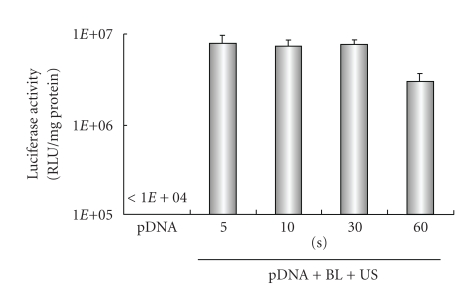
Figure 3.
Effect of ultrasound exposure time on transfection with bubble liposomes into HFLS. pDNA (pCMV-Luc) and BL were mixed together with culture medium and added to the HFLS. The cells were immediately exposed to US (intensity, 2.5 W/cm2; US exposure time, 5–60 sec.). The cells were washed and cultured for 2 days, and then luciferase activity was determined. All data are shown as the mean ± SD (n = 4). BL: bubble liposomes; US: ultrasound exposure.

