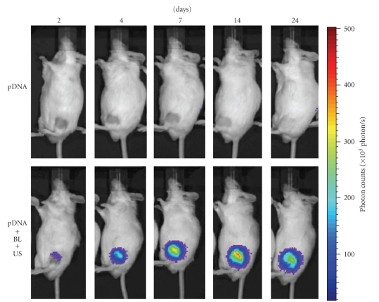
Figure 6.
In vivo luciferase imaging. A suspension of pDNA and BL was injected into the knee joint of the mice, and US exposure (frequency, 1 MHz; duty, 50%; intensity, 2 W/cm2; time, 60 sec) was immediately applied at the injection site. Luciferase expressions after transfection into the joint treated with pDNA, or pDNA plus BL plus US exposure were observed with an in vivo luciferase imaging system for 2–24 days.

