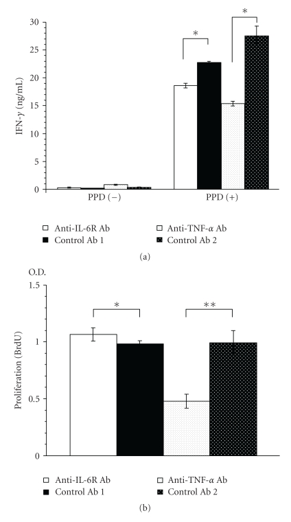
Figure 5.
IFN-γ production and proliferation of spleen cells obtained 4 weeks after TB infection from Ab-treated mice in response to TB antigens (PPD). (a) Spleen cells from each group (n = 5) were pooled and cultured with 20 μg of PPD or TB antigens per mL for 48 h, and then IFN-γ in the culture supernatant was measured. Results are the mean ± S.E.M of triplicate culture. *P < .01 versus corresponding control by Student's t-test. (b) Spleen cells were incubated in the presence of PPD, and BrdU incorporation in the nucleus was measured as O.D. ***(optical density at 340 nm) as an indicator of proliferation. Results are the mean ± S.E.M. of triplicate samples from 5 mice per group. *P < .05 and **P < .01 versus corresponding control by Student's t-test.

