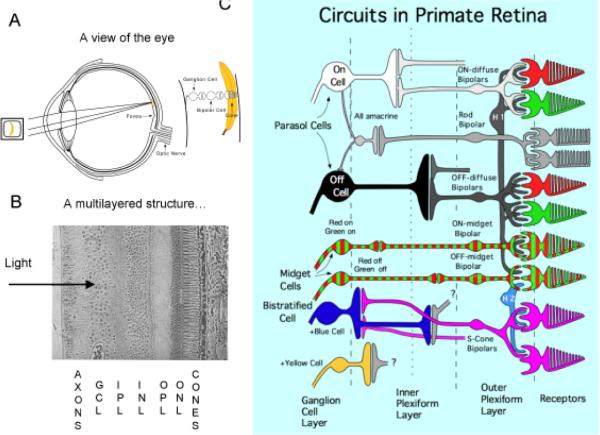
Fig. 7.
Current view of the retina. A. A cross-section of the eye; images are focused on the receptor layer and signals pass through bipolar cells to the ganglion cells, which send axons to the brain via the optic nerve. B The retina is highly laminated structure. The cone receptors are at back, and the Outer Nuclear Layer (ONL) contains the cell bodies of cones and horizontal cells. Outer Plexiform Layer (OPL) contains a synaptic network between cones, bipolar cells and horizontal cells. The Inner Nuclear Layer (INL) contains cell bodies of bipolar cells and amacrine cells, and the Inner Plexiform Layer (OPL) contains a synaptic network between bipolar cells, amacrine cellls and ganglion cells. The Ganglion Cell Layer (GCL) contains ganglion cell bodies which send their axons to the brain. C. A wiring diagram of the primate retina. There are three main cell systems which send axons to the cortex via the lateral geniculate nucleus. Each receive input from the cones, and signals pass through specialized bipolar cells to the ganglion cells. Amacrine and horizontal cells are interneurones which aid local processing of signals. There is an achromatic, luminance pathway beginning in the parasol ganglion cells, a red-green pathway beginning in the midget ganglion cells and a blue-yellow pathway made up of small bistratified cells and another type. These color opponent systems receive opponent input from the different cones, but the color channels do not map perfectly onto the psychophysical opponent systems.

