
Figure 3.
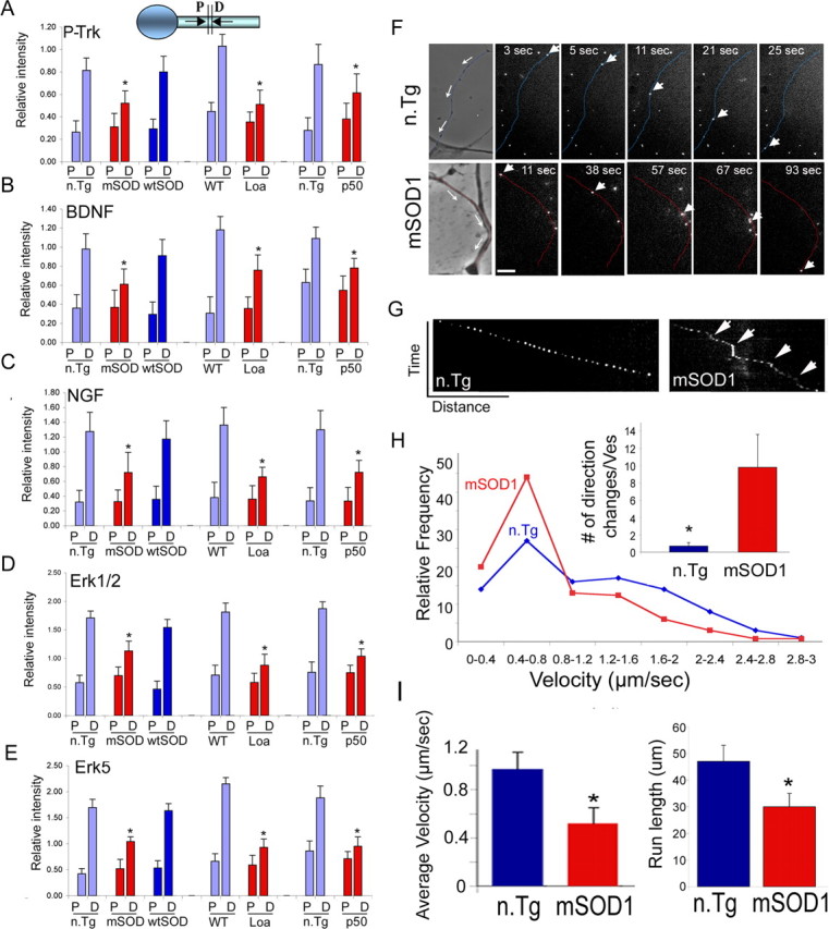
Inhibition in the retrograde transport of trophic factors. A–E, Double ligation assays of sciatic nerves of 85-d-old SOD1G93A (mSOD), 6-month Loa, and 1-year Tgdynamitin overexpressing mice (p50) and age-matched control mice were performed, and segments immediately proximal (P) and distal (D) to the ligation site were subjected to Western blot analysis. Significant inhibition of the retrograde transport of survival factors such as P-Trk (A), BDNF (B), NGF (C), Erk1/2 (D), and Erk5 (E) was observed in all three models. *p < 0.01. F, NGF-Qdot transport was imaged in DRG neurons cultured from 85-d-old mSOD1 and n.Tg control mice. The arrows in the phase image show the direction of movement, the arrowheads track representative Q-dots, and the neurites have been pseudocolored in blue (n.Tg) or red (mSOD). Scale bar, 5 μm. G, A representative kymograph shows more pauses (arrows) in the transport of NGF-Qdots in neurons expressing mSOD1. H, The relative frequency distribution for instantaneous velocities (n > 120) shows a significant shift to lower velocities. The Qdot-NGF changes direction of movement many more times in neurons expressing mSOD1 compared with control (*p < 0.01). I, The average retrograde velocity of NGF-Qdots and the total run length were significantly decreased (n ≥ 5; *p < 0.01) (average velocity ± SEM).