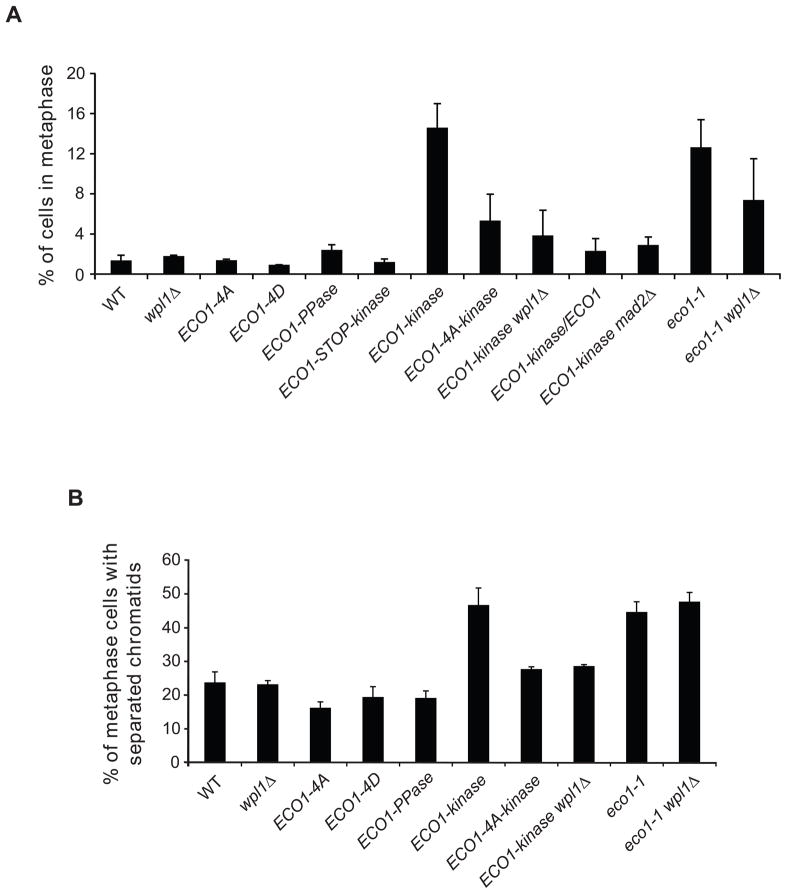
Figure 2. Phosphorylation inhibits the cohesion establishment function of Eco1.
(A) Cells of the indicated genotype were grown asynchronously at room temperature and examined by immunofluorescence microscopy, staining for tubulin and DAPI. Cells were scored for cell cycle stage, defining a metaphase cell as large-budded with a single DNA mass and short bipolar spindles. Over 200 cells were counted in two independent experiments; error bars represent the standard error of the mean (SEM).
(B) Strains containing a GFP-tagged URA3 locus were arrested in metaphase at 30°C using nocodazole, and scored for one GFP focus (maintenance of cohesion) or two foci (loss of cohesion). Over 200 metaphase-arrested cells were counted, and an average was calculated from two or three independent experiments; error bars represent SEM.

