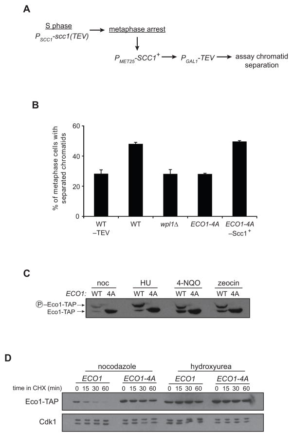
Figure 6. Unphosphorylated Eco1 can establish cohesion after S phase, and Eco1 is stabilized in the presence of DNA damage.
(A) Experimental setup for measurement of cohesion establishment in metaphase. Cohesion in these strains is established normally in S phase with Scc1(TEV). Cultures are arrested in metaphase, wild-type Scc1 is induced for 1 h, TEV is induced for 2 h, and cohesion of GFP-tagged chromatids is analyzed by microscopy.
(B) Cells were treated as indicated in panel A, and separation of sister chromatids was measured in over 200 metaphase cells in each of two experiments; bars indicate mean +/− SEM. Controls were included in which S-phase cohesion was not cleaved (−TEV) or wild-type Scc1 was not induced in metaphase (−Scc1+). The difference between WT and ECO1-4A is statistically significant (P=0.004); for all other differences, P≤0.02.
(C) Cultures of ECO1-TAP (WT) or ECO1-4A-TAP (4A) were treated with nocodazole (noc), hydroxyurea (HU), 4-nitroquinoline 1-oxide (4-NQO), or zeocin for 3 h. Lysates were analyzed by western blotting, using Phos-tag reagent to measure phosphorylation state.
(D) ECO1-TAP and ECO1-4A-TAP strains were arrested by treatment with nocodazole or hydroxyurea, and Eco1 half-life was analyzed by addition of CHX.

