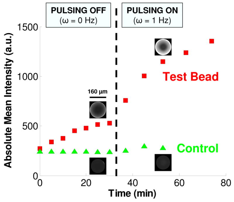
Figure 2.

The emission intensities of a streptavidin-coated, test bead and a control (unfunctionalized) bead as functions of time. During the first 30 min, the beads are not pulsed. Pulsing at 1 Hz commences at 30 min and is maintained for the duration of the experiment. The micrographs for each bead are at t = 25 min and t = 53 min. The concentration of QDots in the buffer is 100 nM. Images are taken with a 20× objective at 2 ms exposure. The conduit is 125 μm tall.
