
Figure 1.
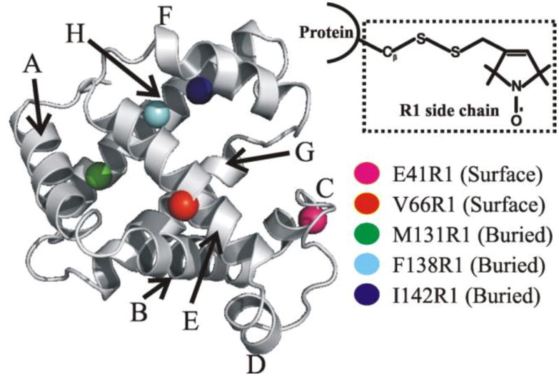
Structure of sperm whale myoglobin (PDB: 2mbw54; the heme prosthetic group is omitted for clarity). The eight α-helices are labeled and the five sites analyzed in this work are highlighted. The inset shows the R1 spin label generated via reaction of a methanethiosulfonate reagent with a cysteine side chain thiol. Image created with PyMOL (version 0.99; DeLano Scientific, San Carlos, CA).