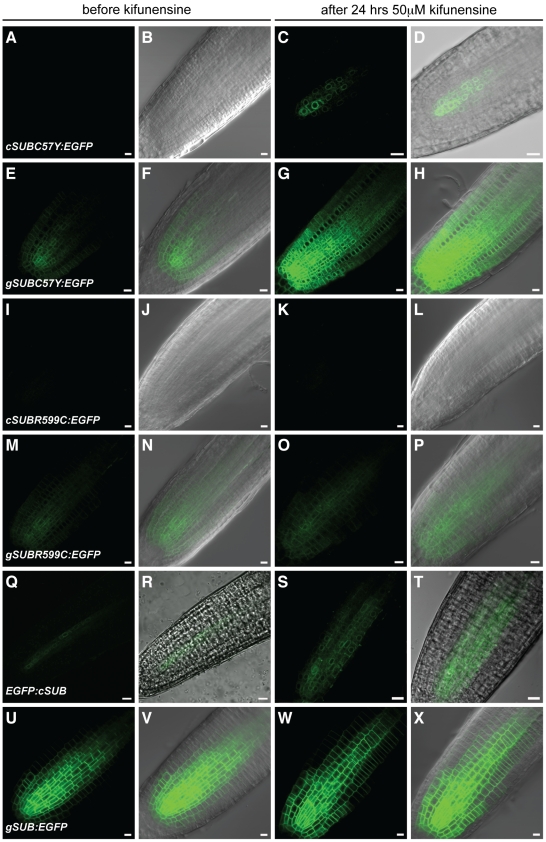
Figure 10. Effects of kifunensine treatments on the expression of different SUB::c/gSUB:EGFP reporters.
Live confocal microscopy images from roots were generated using 4-day old Arabidopsis seedlings (Ler) carrying different SUB:EGFP reporters. Signals from the EGFP channel are shown in green. Differential interference contrast (DIC) or brightfield photomicrographs are shown to outline root tissue (B, D, F, H, J, L, N, P, R, T, V, X). The same root before (A–B, E–F, I–J, M–N, Q–R, U–V) and after (C–D, G–H, K–L, O–P, S–T, W–X) 24-hrs treatment with 50 µM kifunensine. (A–D) SUB::cSUBC57Y:EGFP. Signal becomes detectable upon kifunensine treatment. Note ER-like pattern (compare with Figure 9S–T). (E–H) SUB::gSUBC57Y:EGFP. Signal becomes stronger upon kifunensine treatment. (I–L) SUB::cSUBR599C:EGFP. Absence of signal, irrespective of kifunensine treatment. (M–P) SUB::gSUBR599C:EGFP. Signal is easily detectable and not noticeably influenced by kifunensine treatment. (Q–T) SUB::EGFP:cSUB. Note the ER-like pattern (compare with Figure 9S–T). No change in signal intensity was observed upon kifunensine treatment. (U–X) SUB::gSUB:EGFP. The reporter signal does not change detectably upon treatment with kifunensine. Scale bars: 10 µm.

